Microbiomes are all the scientific rage, even in art conservation, where studying the microbial species that congregate on works of art may lead to new ways to slow down the deterioration of priceless aging artwork, as well as potentially unmask counterfeits. For instance, scientists have analyzed the microbes found on seven of Leonardo da Vinci's drawings, according to a recent paper published in the journal Frontiers in Microbiology. And back in March, scientists at the J. Craig Venter Institute (JCVI) collected and analyzed swabs taken from centuries-old art in a private collection housed in Florence, Italy, and published their findings in the journal Microbial Ecology.
The researchers behind the earlier March paper were JCVI geneticists who collaborated with the Leonardo da Vinci DNA Project in France. The work built on a prior study looking for microbial signatures and possible geographic patterns in hairs collected from people in the District of Columbia and San Diego, California. They concluded from that analysis that microbes could be a useful geographic signature.
For the March study, the JCVI geneticists took swabs of microbes from Renaissance-style pieces and confirmed the presence of so-called "oxidase positive" microbes on painted wood and canvas surfaces. These microbes munch on the compounds found in paint, glue, and cellulose (found in paper, canvas, and wood), in turn producing water or hydrogen peroxide as byproducts.
"Such byproducts are likely to influence the presence of mold and the overall rate of deterioration," the authors noted in their paper. "Though prior studies have attempted to characterize the microbial composition associated with artwork decay, our results summarize the first large-scale genomics-based study to understand the microbial communities associated with aging artwork."
As an added bonus, they found they could discern between microbial populations on different types of materials. Specifically, stone and marble art fostered more diverse populations than paintings, possibly due to the "porous nature of stone and marble harboring additional organisms and potentially moisture and nutrients, along with the likelihood of biofilm formation," they wrote. Oil paintings offered more meager nutrients for microbes to metabolize, by contrast.
The authors acknowledged the small sample size, but they nonetheless concluded that microbial signatures could be used to differentiate artwork according to the materials used. As always, more research is needed. "Of particular interest would be the presence and activity of oil-degrading enzymes," the authors wrote. "Such approaches will lead to fully understanding which organism(s) are responsible for the rapid decay of artwork while potentially using this information to target these organisms to prevent degradation."
Swabbing Renaissance art
-
Collage of the various artworks sampled for a March paper by geneticists at the J. Craig Venter Institute. Circles indicate swabbed areas on each sample artworkJCVI
-
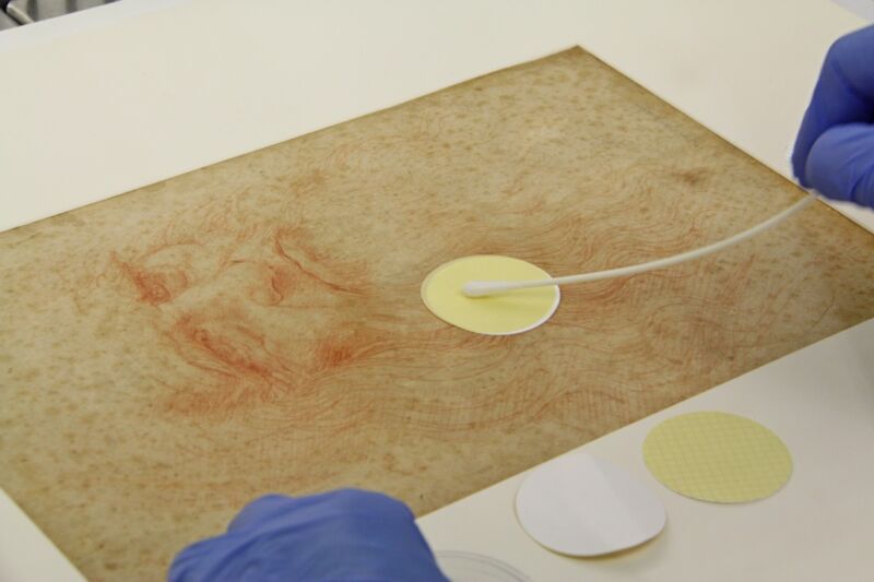
Manolito G. Torralba and colleagues used small, dry polyester swabs to gently collect microbes from centuries-old, Renaissance-style art in a private collector's home in Florence, Italy.Jesse Ausubel
Guadalupe Piñar and her team at the University of Natural Resources and Life Sciences in Vienna, Austria, collaborated with conservators from the Instituto Centrale per la Patologa degli Archivi e del Libro (ICPAL) for the microbiome analysis of the Leonardo da Vinci drawings. Last year, Piñar et al. relied upon microbiome analysis to study the storage conditions of three statues retrieved from smugglers, as well as pinpointing their possible geographical origins. Earlier this year, they analyzed the microbiome of 1,000-year-old parchments, from which they were able to deduce the animals whose skins were used to make the parchments.
For this latest paper, Piñar's team turned to a third-generation sequencing method known as Nanopore, which uses protein nanopores embedded in a polymer membrane for sequencing. It comes with a portable, pocket-size sensing device, the MinION, making it ideal for cultural heritage studies. For the Leonardo drawings, Piñar et al. combined the Nanopore sequencing with a whole-genome-amplification protocol.
The ICPAL conservators used a delicate, noninvasive microaspiration (i.e., filtering suction) sampling method to collect dust particles, microbial cells, and other debris from small surface areas on both the recto and verso of each drawing. Then the DNA was extracted, amplified, and sequenced by the Austrian team. They used optical microscopy to image features of interest in all seven drawings and scanning electron microscopy (SEM) to analyze all the micro-objects gathered from the drawings.
A “biological pedigree”
-
The drawings by Leonardo da Vinci analyzed in a study by Austrian and Italian scientists.Guadalupe Piñar et al.
-
Sampling microbiome from Leonardo da Vinci's Studio di panneggio per una figura inginocchiata (ca. 1475).Guadalupe Piñar et al.
-
Leonardo da Vinci's Uomo della Bitta.Guadalupe Piñar et al.
-
Insect droppings appear as waxy brown incrustations on the fibers of drawing L4 (Studi delle gambe anteriori di un cavallo).Guadalupe Piñar et al.
-
SEM images of the surface of a membrane used to sample the surface of the drawing L3 (Nudi per la battaglia di Anghiari).Guadalupe Piñar et al.
Each drawing had its own unique microbiome—an "independent molecular profile or biological pedigree." But Piñar et al. were surprised to find that, overall, bacteria dominated fungi in the drawings' microbiomes, contradicting widespread belief that fungi would be more dominant, given their higher potential to colonize on paper-based works. The researchers detected no visible biodeterioration on the drawing, apart from foxing stains (small yellow-brown spots or blotches).
Much of those bacteria are typically found in human microbiomes, suggesting they found their way onto the drawings while being handled during restoration—although one could speculate about whether it came from the artist himself. (The authors note that bacteria in dust could "remain in suspension" for long periods.) Other bacteria were typical of insect microbiomes and may have been introduced long ago by flies depositing excrement on the drawings. Those droppings showed up under imaging analysis as waxy brown incrustations in the fibers.
The Austrian/Italian team was unable to conclude definitively whether any of the microbial contaminants date back to Leonardo's time. It seems far more likely that the human microbial elements are due to more recent restoration work. This is most likely to be the case for the drawing designated L4 (Studi delle gambe anteriori di un cavallo) in particular, which showed the least biodiversity and the heaviest contamination from human DNA. The authors hypothesize further that the recipe Leonardo used—"a preparatory layer made with powdered calcinated chicken bones, white lead, indigo... mixed with animal gelatin"—may have interfered with the preservation of the microbiome of L4, so only recent DNA remained.
But Piñar insists that being able to track this kind of data is nonetheless highly valuable. "The sensitivity of the Nanopore sequencing method offers a great tool for the monitoring of objects of art. It allows the assessment of the microbiomes and the visualization of its variations due to detrimental situations," she said. "This can be used as a bio-archive of the objects' history, providing a kind of fingerprint for current and future comparisons."
DOI: Microbial Ecology, 2020. 10.1073/pnas.1802831115
DOI: Frontiers in Microbiology, 2020. 10.1007/s00248-020-01504-x (About DOIs).
reader comments
44